Elastography
Elastography is a new and promising technique for the detection of thyroid malignancy. This technology has the potential to become part of the standard thyroid ultrasound examination. We are very excited to offer this technology in our office. Elastography evaluates tissue stiffness and can be very useful in the prediction of malignancy.
ELASTOGRAPHY: SEEING THROUGH CANCERSM
Advancing the art of thyroid cancer detection
Elastography is a new and promising technique for the detection of thyroid malignancy. This technology has the potential to become part of the standard thyroid ultrasound examination. We are very excited to offer this technology in our office elastography evaluates tissue stiffness and can be very useful in the prediction of malignancy. It is known that malignant lesions tend to be stiffer, or harder, than the surrounding tissue. Benign lesions tend to have the same or less stiffness then the surrounding tissue. Conventional Ultrasound exam is not capable of detecting such abnormality that could otherwise be missed.
There are two general ultrasonic methods for determining elastography, strain wave and shear wave.
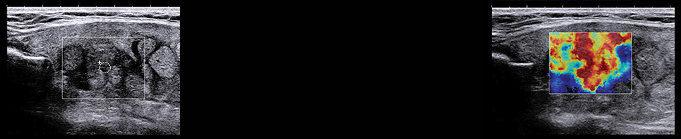
Strain elastography: This method of elastography requires manual compression of the tissue being visualized. This manual compression, by the ultrasound technician, will move the underlying tissue. The more elastic the tissue is the more movement is experienced. The movement of the tissue is then measured and visualized on a color map.
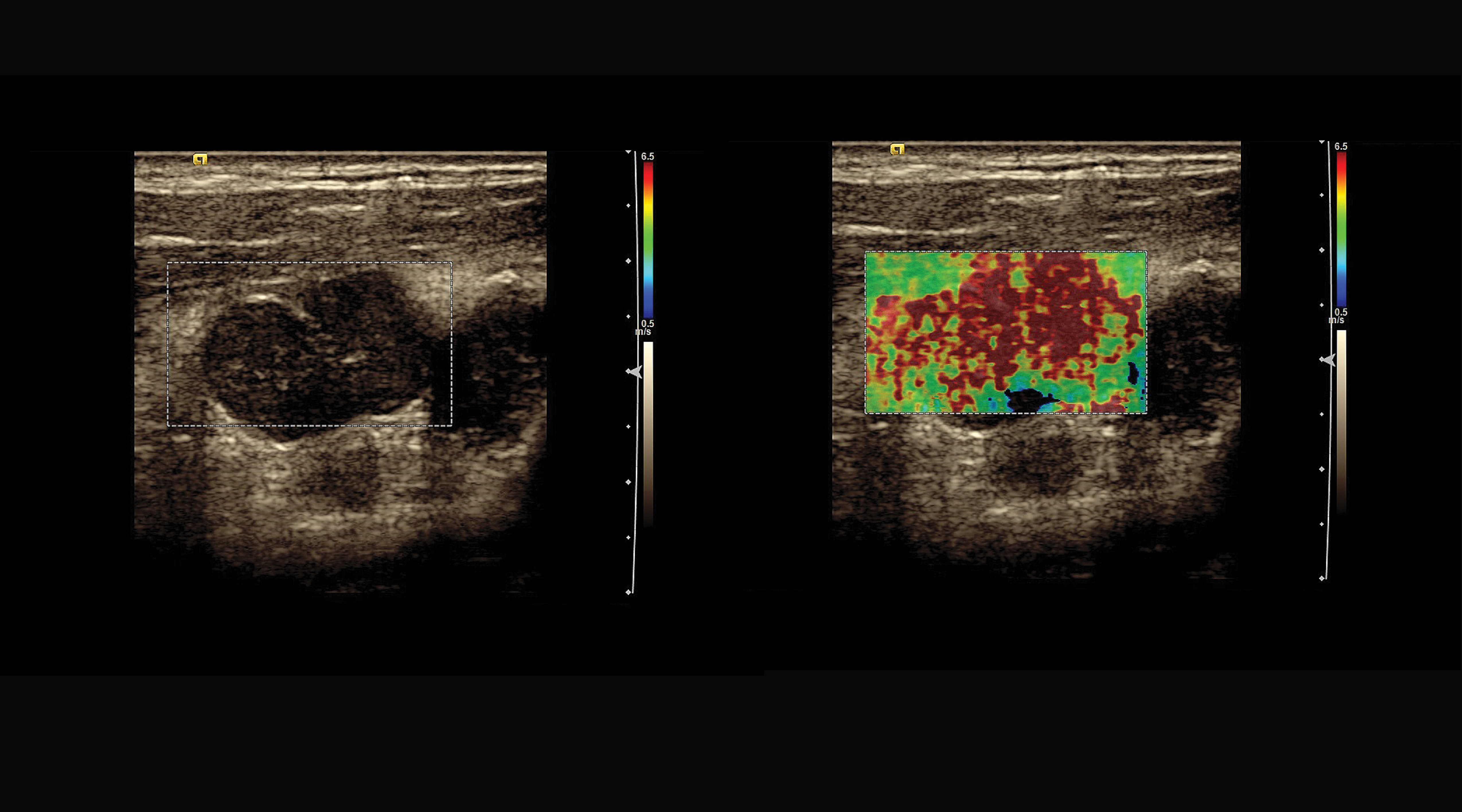
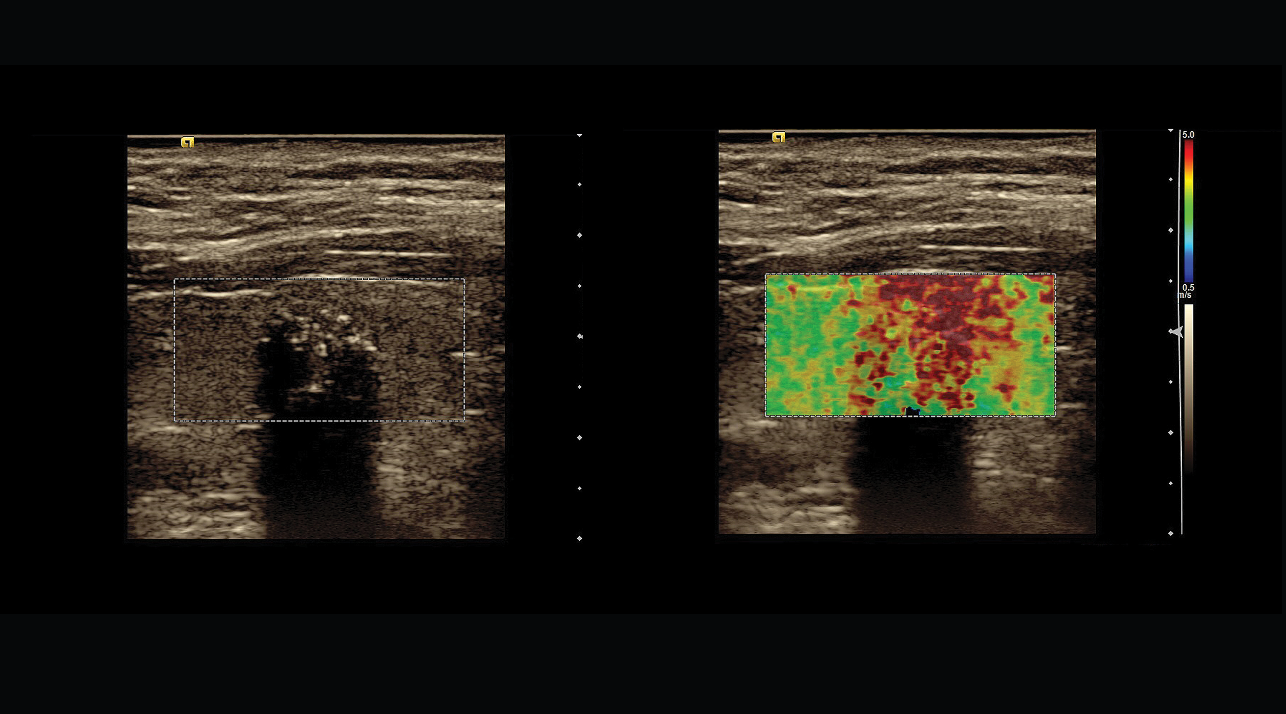
Shear Wave elastography: This method of elastography uses a patented technology called “Sonic Touch” to create a shear wave that travels through the tissue. This movement creates sound waves that will cause a slight displacement of the tissue much like ripples in a pond. The stiffer the tissue is the faster the shear wave will travel through the tissue. The wave is tracked, measured, and given a numerical value of the shear wave speed.
The tissue stiffness is then depicted on a color map. The different colors represent the different tissue densities. Our color map uses the color red to represent the hardest tissue and the color blue to represent the softest tissue.
SonogenicSM nodules are those with high probability for malignancy based on b-mode ultrasound features, strain elastography and shear wave elastography.
We believe that this technology, in conjunction with other ultrasound features can vastly improve the diagnosis and treatment of thyroid nodules and thyroid cancer. This technology can improve our ability to predict thyroid malignancy prior to Fine Needle Aspiration Biopsy (FNAB), when performing ultrasound exam. Elastography may reduce the number of thyroid nodule FNAB for lesions with low probability for malignancy based on ultrasound features. Additional prospective studies are needed. Our prospective elastography study is ongoing.
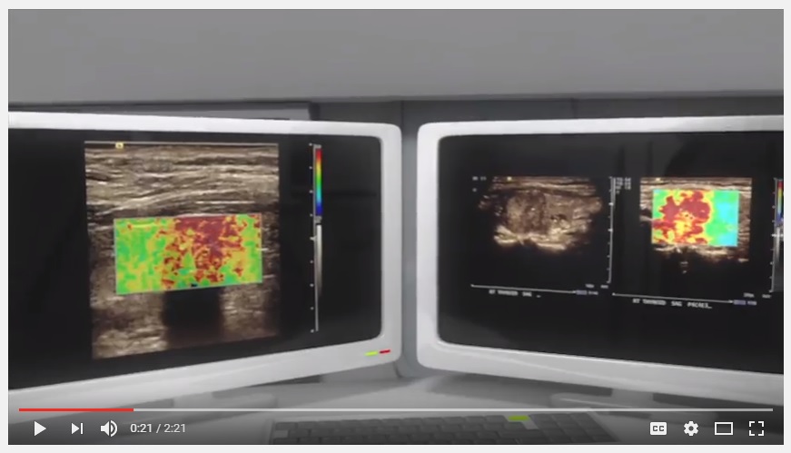
Wilmington Endocrinology and Thyroid Media are pleased to present their latest animation: Thyroid Elastography! This video shows viewers the use of ultrasound and elastography exam in assessing the risk for malignancy in thyroid nodules. You can watch the video by clicking HERE.