
Connecting Tomorrow’s Technology with Today
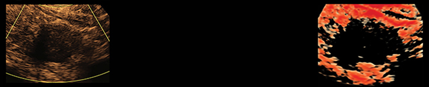
Ultrasound technology is being used to visualize the thyroid gland and detect thyroid malignancy. Ultrasound technology works by sending out high frequency sound waves which pass through the body and are reflected back. The reflected back echoes are then analyzed and built into a picture of the visualized body part. Ultrasound technology has been utilized for over 30 years. Ultrasound technology is useful in obstetrics, gynecology, orthopedics, internal medicine, emergency medicine, cardiology, and in many other fields of medicine. In our office, we utilize ultrasound technology on a daily basis for the visualization of the thyroid gland and detection of thyroid cancer. In addition, we use ultrasound to diagnose parathyroid adenomas.
Ultrasound technology is used quite often in the field of Endocrinology. It helps to define the shape and size of the thyroid gland and any lesions within the gland. In addition it can help assess the texture and vascularity of the thyroid tissue. Ultrasound also allows the operator to detect the presence of thyroid nodules that may or may not be malignant. There are certain sonographic features that are useful in differentiating benign versus malignant lesions. Ultrasound technology is also being utilized during Fine Needle Aspiration biopsy of thyroid lesions. The incidence of thyroid malignancy is increasing more rapidly than that of any other malignancy.
The current 2D technology has several limitations which can lead to misdiagnosing thyroid cancer. This makes it difficult for the physician to be absolutely certain of the proper diagnosis.
Ultrasound machines are becoming more advanced. Some high end machines are now offering 3D and 4D ultrasound technology. 2D ultrasound sends out the sound wave in one direction and the echo returns in one direction. The 2D ultrasound shows everything in 2 dimensions on one plane, sagittal and transverse. 3D ultrasound sends out sound waves in multiple different angles and the echoes return in different angles. The 3D ultrasound introduces a second plane with the addition of a third dimension, sagittal, transverse, and coronal. There is no coronal view in 2D ultrasound. 4D ultrasound is 3D ultrasound in motion. The fourth dimension added is that of time.
The initial difference in a 2D and 3D image is like looking at a loaf of bread. A 2D image is one slice of bread, although looking at it you may think you are looking at the whole loaf of bread. In this case, you are actually only looking at one slice of the thyroid lobe. A 3D image contains the whole loaf of bread, or in this case the whole thyroid lobe. The 2D image or slice of bread does not portray the “volume” of the loaf of bread, whereas the 3D image easily allows you to visualize the total volume of the entire loaf of bread. This “volume” of information can be used as multiple slices of the thyroid but can also be turned and rotated to display individual layers or “slices” from multiple angles. It allows you to see the accurate shape of the nodule, the way it is seen in reality.
3D ultrasound technology has been mainly used in the fields of obstetrics and gynecology. It has been found, that 3D technology is more accurate for visualizing cleft lip, cleft palates, and other fetal abnormalities than 2D technology. It is used in the detection of ovarian cancer, endometrial cancer and cervical cancer[1]. It is starting to be used for transesophageal echocardiography [6] and carotid stenosis[5]. We have utilized this 3D technology in our office to detect malignant thyroid nodules referred to as 3DTHYROID and to help with needle guidance for FNA biopsy.
3D ultrasound allows for more in-depth and focused view of the thyroid nodule. It allows the physician to view the actual shape of the nodule and assess for any irregularities. These irregularities are worrisome features that could increase the patient’s risk for malignancy. They may be missed with the conventional 2D ultrasound.
3D ultrasound allows you to see the shape and borders of the thyroid nodule more accurately. It also allows you to assess the volume of the lesion precisely as opposed to current 2D technology. This technology enables you to define therelation to the nodule’s proximity to the thyroid capsule and possible invasion of the thyroid capsule. The invasion of the thyroid capsule is indicative of malignancy. This is also very important in assessing the potential for metastatic disease. This may become very useful to the surgeons preoperatively. The 3D technology also allows the structures that are attached or adjacent, to the thyroid gland to be seen more in depth. These structures include parathyroid adenomas, ectopic thyroid tissue, possible malignant lymph nodes, and blood vessels that should be avoided during Fine Needle Aspiration Biopsy. 3D ultrasound allows you to see if these structures are adjacent to the thyroid gland versus within the thyroid gland.
Fine Needle Aspiration biopsy (FNA biopsy) of thyroid lesions, with the 2D ultrasound shows the nodule and needle in 2 dimensions in one plane. This may falsely show the needle within the lesion when the needle is actually adjacent to the lesion, (superior or inferior). This worry is taken away with the introduction of the third dimension and second plane. It allows you to know that the needle is, in fact, inside the lesion. This is particularly helpful when the lesion in questions is deep in the neck. The 3D ultrasound also allows you to rotate the image and see all the borders. The national average for false negative Fine Needle Aspiration biopsy pathology report is 5%.[2,3,4] A false negative report, is one where the pathologist states that there is no thyroid cancer, when in reality thyroid cancer exists. The elevated false negative rate is mainly caused by visual misperception caused by current 2D technology. 3D ultrasound can significantly decrease the rate of false negative reports, by eliminating the possibility of the needle being inferior or superior to the lesion during FNA biopsy.
In summary, the incidence of thyroid cancer is increasing more rapidly than that of any other malignancy. 3D and 4D ultrasound technology are superior to conventional 2D technology for distinguishing benign from malignant thyroid nodules. 3D ultrasound should be utilized more frequently in the assessment and treatment of patients with thyroid disease. 3D and 4D ultrasound technology can:
1. Define the shape and size of a thyroid lesion (nodule) more accurately. It identifies possible irregular shape and margins more precisely. Irregular shape is an independent risk factor for thyroid malignancy.
2. Provides useful information in measuring the volume of thyroid nodules.
3. Reduce the rate for False Negative pathology reports for Fine Needle Aspiration Biopsy.
Our own clinical practice is a leader in the diagnosis of thyroid cancer. [3] I am currently using the new 3D technology to identify more thyroid malignancies, that are smaller (5-9 mm) with irregular shape and subcapsular location, than would be possible with 2D technology alone. My on-going prospective thyroid nodule study (started in January 2010) is examining further advantages of the use of 3D ultrasound technology. As with all other malignancies, early detection of thyroid cancer is predicted to improve the patient’s outcome. We believe that 3D technology will be the gold standard for the assessment of patients with thyroid nodules in the near future.
3DTHYROIDSM and 4DTHYROIDSM may enable patients to see as much as the doctor sees.