
Clinical Applications in a Few Words
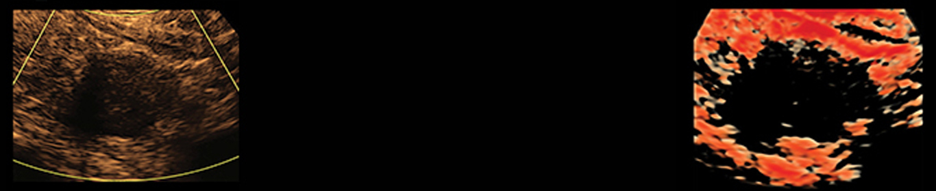
In our office, we utilize ultrasound technology on a daily basis for the visualization of the thyroid gland and detection of thyroid cancer. In addition, we use ultrasound to diagnose parathyroid adenomas.
We have utilized this 3D technology, referred to as 3DTHYROID®, in our office to detect malignant thyroid nodules and to help with needle guidance for FNA biopsy.
3D ultrasound allows for more in-depth and focused view of the thyroid nodule. It allows the physician to view the actual shape of the nodule and assess for any irregularities.
3D ultrasound allows you to see the shape and borders of the thyroid nodule more accurately. It also allows you to assess the volume of the lesion precisely as opposed to current 2D technology. This is also very important in assessing the potential for metastatic disease and may become very useful to the surgeons preoperatively. 3D ultrasound has the potential to be useful for routine assessment and treatment of patients with thyroid disease.
With 3D ultrasound technology, viewing of the margins of thyroid lesions are more defined when compared with 2D ultrasound image.
The accuracy of the current 2D measurement is limited, particularly for the lesions with irregular margins or shape. 3DTHYROID® technology uses 3D volume measurement that may provide more useful information about these lesions.
The increase in the usefulness of 3D ultrasound is directly related to the limitations of 2D technology. With the current conventional ultrasound, the image provided is a two-dimensional, yet the anatomy being visualized is three-dimensional.
Real-time capacity is not generally available with three-dimensional ultrasound. (Real time three-dimensional ultrasound is also known as 4D ultrasound.) Whereas 3D ultrasound is a static display of the various reformatting techniques based on the acquisition of a static volume, 4D ultrasound displays a continuously updated and newly acquired volume in any rendering modality creating the impression of a moving structure.
We believe that 3D technology will be the gold standard for the assessment of patients with thyroid nodules in the near future.