Case of the Month!
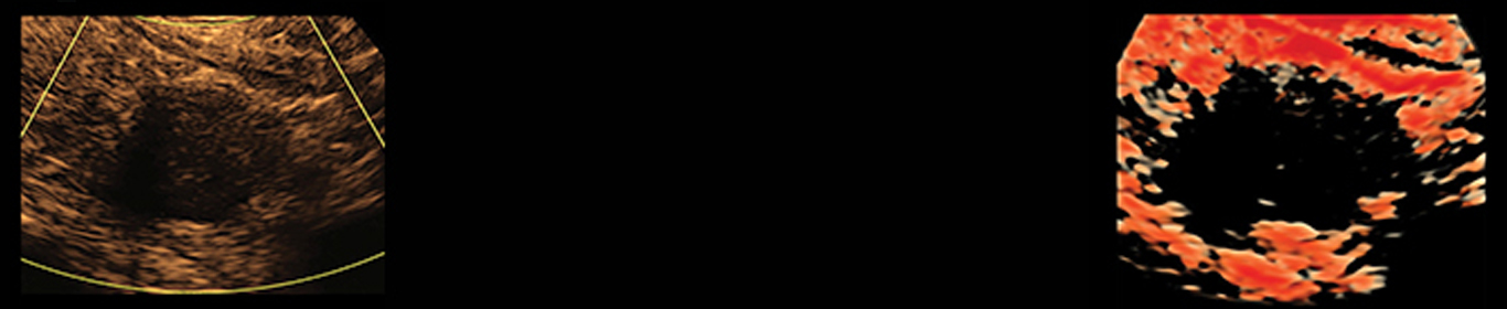
What was your guess for the diagnosis of this 5mm hypoechoic lesion?
Answer: It’s a parathyroid adenoma!
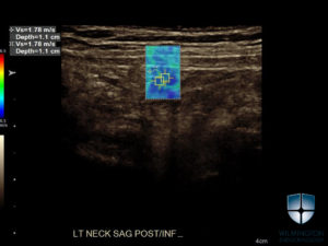
Our latest study examined the value of Shear Wave elastography in identifying and diagnosing parathyroid adenomas.
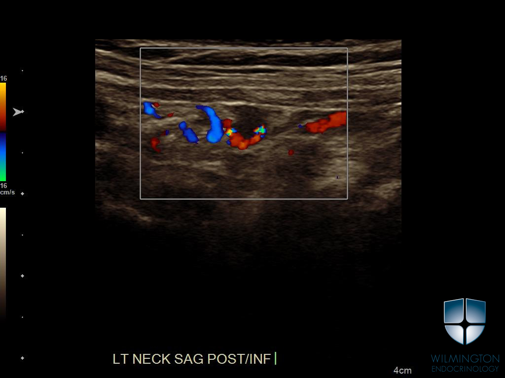
The results demonstrated the median (or average) shear wave velocity for the parathyroid adenoma was 0.76 m/s lower than the surrounding thyroid tissue. In addition, parathyroid adenomas appear to have a more homogenous texture. Vascularity pattern was also found to be helpful as the blood vessel source in the parathyroid contributed to determining whether a mass surrounding the parathyroid was an adenoma or a lymph node. Adenomas have a blood vessel, or polar vessel sign, whereas lymph nodes do not.
While all adenomas in this study were confirmed by MIBI, fine-needle aspiration, or surgical resection, the information gathered is important because adding VTIQ-generated shear wave elastography to basic ultrasound exam could lower the necessity of these tests. Knowledge of the shear wave velocity for a specific tissue can help in the identification of the tissue and lend to the diagnosis of a patient using ultrasound techniques rather than invasive procedures or costly scans.
You can read the full-text of this study HERE, or check out our PUBLICATIONS page to see all of our research.